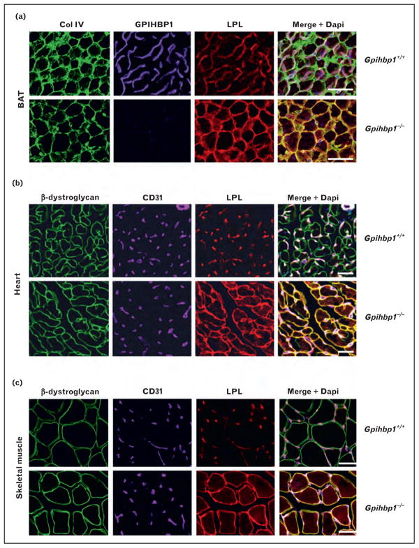
FIGURE 1.
Mislocalization of LPL in tissues of Gpihbp1−/− mice. (a) Confocal microscopy showing the binding of GPIHBP1-specific antibodies, LPL-specific antibodies, and collagen IV-specific antibodies to brown adipose tissue (BAT) from wild-type and Gpihbp1−/− mice. Scale bars, 100 μm. (b, c) Confocal microscopy showing the binding of CD31-specific antibodies, LPL-specific antibodies, and β-dystroglycan-specific antibodies to heart (b) and skeletal muscle (c) of wild-type and Gpihbp1−/− mice. The scale bars show a distance of 100 μm (skeletal muscle) or 50 μm (heart). Reproduced with permission from [29■■].

