Figure 2.
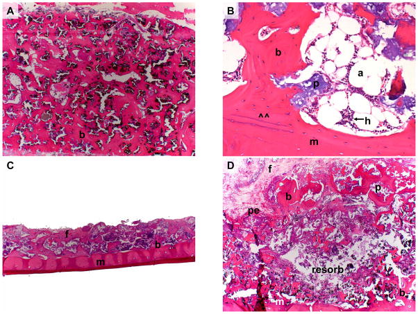
Figure 2A: BMSC-containing transplant (Animal 2) 17 months post-operatively. Note extensive cortico-cancellous bone (b) formed throughout the entire thickness of the transplant.
Figure 2B: BMSC-containing transplant (Animal 3) 19 months post-operatively. Note extensive cortico-cancellous bone encircling hematopoiesis (h) and adipocytes (a). Newly formed bone has formed a union (at ^^) with the underlying mandible (m).
Figure 2C: BMSC-free transplant (Animal 2) 17 months post-operatively. Note that residual transplant has minimal volume and that much of the original HA/TCP scaffold has been resorbed. Minimal bone (b) and extensive fibrous tissue (f) lay on the mandible (m).
Figure 2D: BMSC-free transplant (Animal 3) 19 months post-operatively. Note resorption of transplant interior leaving behind minimal tissue (resorb), and minimal residual bone (b) along mandible (m) and periosteum (pe).
(b = bone, f = fibrous connective tissue, p = particle, m = mandible, pe = periosteum, a = adipocyte, h = hematopoiesis), resorb = (resorbed tissue within BMSC-free transplant)
Magnification: 2.5x (A, C, D). Magnification: 20x (B).
Stain: Hematoxylin and eosin; paraffin embedding following demineralization (A–D)

