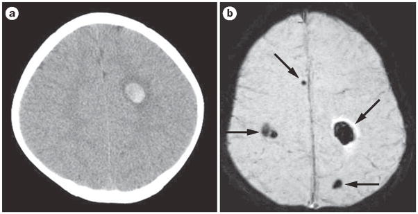
Figure 2.
Acute hemorrhagic stroke and susceptibility-weighted imaging. a | The head CT shows a single, small right frontal acute parenchymal hemorrhage. b | The MRI susceptibility-weighted image shows the right frontal hemorrhage as well as multiple additional hemorrhages not visualized on head CT. This child has multiple cerebral cavernous malformations.

