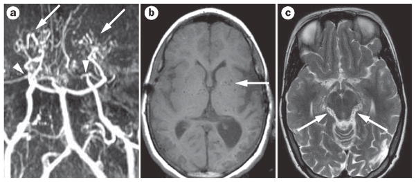
Figure 3.
Idiopathic moyamoya disease. a | Time-of-flight, noncontrast, magnetic resonance angiography shows moyamoya vasculopathy with bilateral occlusion of the internal carotid arteries (arrowheads). Robust lenticulostriate collaterals can also be observed (arrows). b | The lenticulostriate collaterals in moyamoya disease are also seen on the MRI scan, with multiple flow voids piercing the basal ganglia (arrow). c| These moyamoya collateral vessels are also seen as multiple small-flow voids around the brainstem (arrows).

