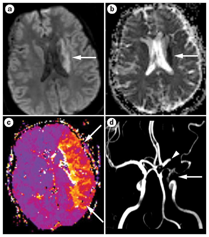
Figure 4.

Acute arterial ischemic stroke with diffusion–perfusion mismatch. a| The MRI reveals a small acute stroke that shows restriction on diffusion-weighted imaging in the periventricular region (arrow). b| The corresponding apparent diffusion coefficient map confirms the occurrence of acute ischemia (arrow). c| The perfusion-weighted image shows a large area of hypoperfusion—basically the entire left middle cerebral artery territory (arrow)—representing diffusion–perfusion mismatch. d| This stroke was caused by embolization of a cardiac thrombus that led to partial occlusion of the internal carotid artery—little flow is seen on magnetic resonance angiography (arrow)—and complete occlusion of the middle cerebral artery (arrowhead).
