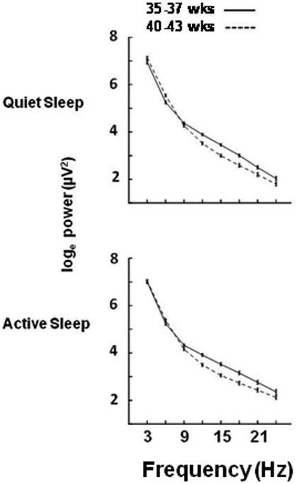
Figure 4.

EEG power (loge, μv2) in the left hemisphere in each frequency band in Quiet (upper panel) and Active (lower panel) Sleep. Data are from prematurely born infants studied from 35 through 37 weeks (N=29) postmenstrual age (solid line) and from 40 through 43 weeks (N=40, dashed line).
