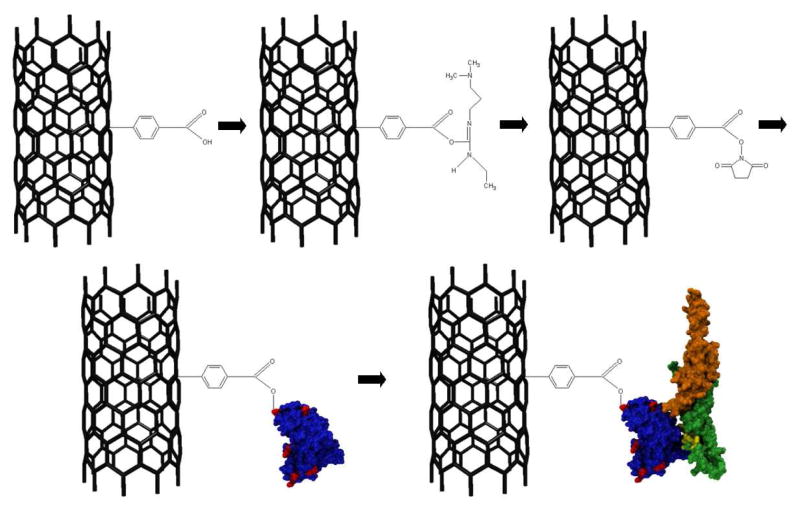
Figure 1. Functionalization scheme for OPN attachment.
First, sp3 hybridized sites are created on the nanotube sidewall by incubation in a diazonium salt solution. The carboxylic acid group is then activated by EDC and stabilized with NHS. ScFv antibody displaces the NHS and forms an amide bond (surface amine-rich lysine residues responsible for this bond are depicted in red), and OPN binds preferentially to the scFv in the detection step. The OPN epitope is shown in yellow, and the C-and N-terminuses are in orange and green respectively.

