Figure 5.

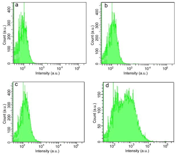
Flow-cytometry measurements of the intensity distributions of cancer cells (MCF-7) labeled via non-specific binding, for (a) control blank sample in the absence of Pdots, (b) PFBT-C2 dots, (c) PFBT-C14 dots, and (d) PFBT-C50 dots.

Flow-cytometry measurements of the intensity distributions of cancer cells (MCF-7) labeled via non-specific binding, for (a) control blank sample in the absence of Pdots, (b) PFBT-C2 dots, (c) PFBT-C14 dots, and (d) PFBT-C50 dots.