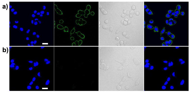
Figure 7.

Fluorescence images of newly synthesized proteins in MCF-7 breast-cancer cells tagged with PFBT-C14A probes. (a) Positive labeling using PFBT-C14A probe. (b) Negative labeling carried out under the same condition as in (a) but in the absence of Cu(I) catalyst. From left to right: blue fluorescence from the nuclear stain Hoechst 34580; green fluorescence images from PFBT-C14A probes; Nomarski (DIC) images; and combined fluorescence images. Scale bars: 20 μm.
