
Figure 6.
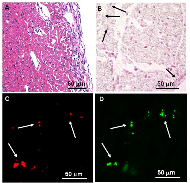
Histopathological analyses of the allograft heart tissue harvested on POD 6: (A) optical micrograph of tissue section stained with H&E for tissue integrity; (B) optical micrograph of tissue section stained with Prussian blue for iron; (C–D) fluorescent microscopic images of double immuno-fluorescent staining with (C) phycoerythrin (PE)-conjugated anti-CD3 and (D) mouse anti-PEG mAb, following treatment with anti-mouse IgG-FITC.