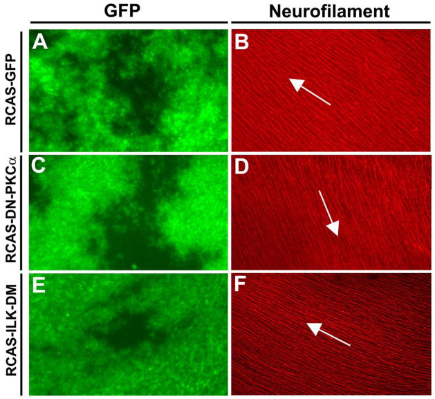
Figure 7.
Intraretinal projection of the RGC axons was not affected by expression of DN-PKCα or ILK-DM. The optic vesicles of the chick embryos were injected with RCAS-GFP, RCAS-DN- PKCα or RCAS-ILK-DM at E1.5. Retinas were harvested at E7, flat-mounted with the ganglion side up, and wide-spread GFP expression was observed (A, C, E). Immunofluorescent staining with an anti-neurofilament antibody (B, D, F ) showed the trajectories of the RGC axons. In all cases, the projection patterns of the RGC axons toward the optic disc appeared normal (some minor aberrant appearance is due to the curvature of the retinal surface). Arrows indicate the direction to the optic disc.

