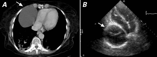
Fig. 2 A) Chest computed tomography (cross-section) and B) transthoracic echocardiography (apical 4-chamber view) show a large, unilocular, thin-walled structure (arrow) of water-density opacity, located lateral to the right atrium and compressing both the right atrium and the inferior vena cava.
