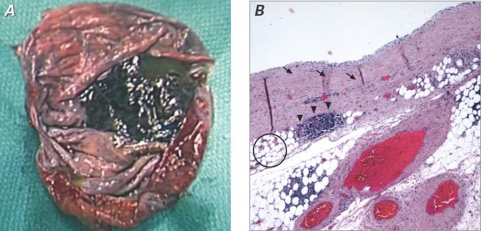
Fig. 4 Pathology specimen. A) Macroscopic view shows an oval-round cystic lesion (8 cm at largest diameter) filled with clear, watery fluid. B) Microscopic view shows that the cystic wall is composed of hypocellular fibroconnective tissue (arrows) with scattered lymphoid aggregates (arrowheads) and a substantial number of fat cells (circle). No malignant features are seen (H & E, orig. ×40).
