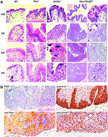
Fig. 3.

Histopathology. (A) Urinary bladders of WT, Ras* and SV40T single transgenic mice and Ras*/SV40T double transgenic mice of newborn (N/B) and 3, 5 and 7 weeks of age were sectioned and stained with hematoxylin and eosin (H&E). Note the normal urothelial morphology in wild-type (WT) mice of all age groups; simple hyperplasia in Ras* mice; severe dysplasia and CIS-like lesions in SV40T mice and in contrast, the rapid progression from dysplasia/CIS-like lesions in N/B to high-grade papillary tumors in Ras*/SV40T double transgenic mice as early as 3 weeks. Magnification: the left column under Ras*/SV40T, ×50; all other panels, ×200. (B) Examination of the basement membrane zone by H&E and antibody staining. Representative images from serial sections of bladders of 5- to 6-week-old Ras*/SV40T double transgenic mice were stained with H&E or with antibodies against keratin 14, pan-Ras or SV40T. Note the absence of tumor cells beyond the basement membrane.
