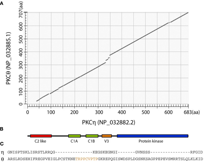
Figure 2.
Comparison of mouse PKCη and PKCθ proteins. (A) Alignment of mouse PKCη and PKCθ was performed using NCBI BLAST program. (B) Cartoon showing the arrangement of known conserved domains that applies to both PKCη and PKCθ proteins. (C) Alignment of PKCη and PKCθ V3 domains. The region identified as important for PKCθ interaction with CD28 (Kong et al., 2011) is highlighted in orange.

