Fig. 1.
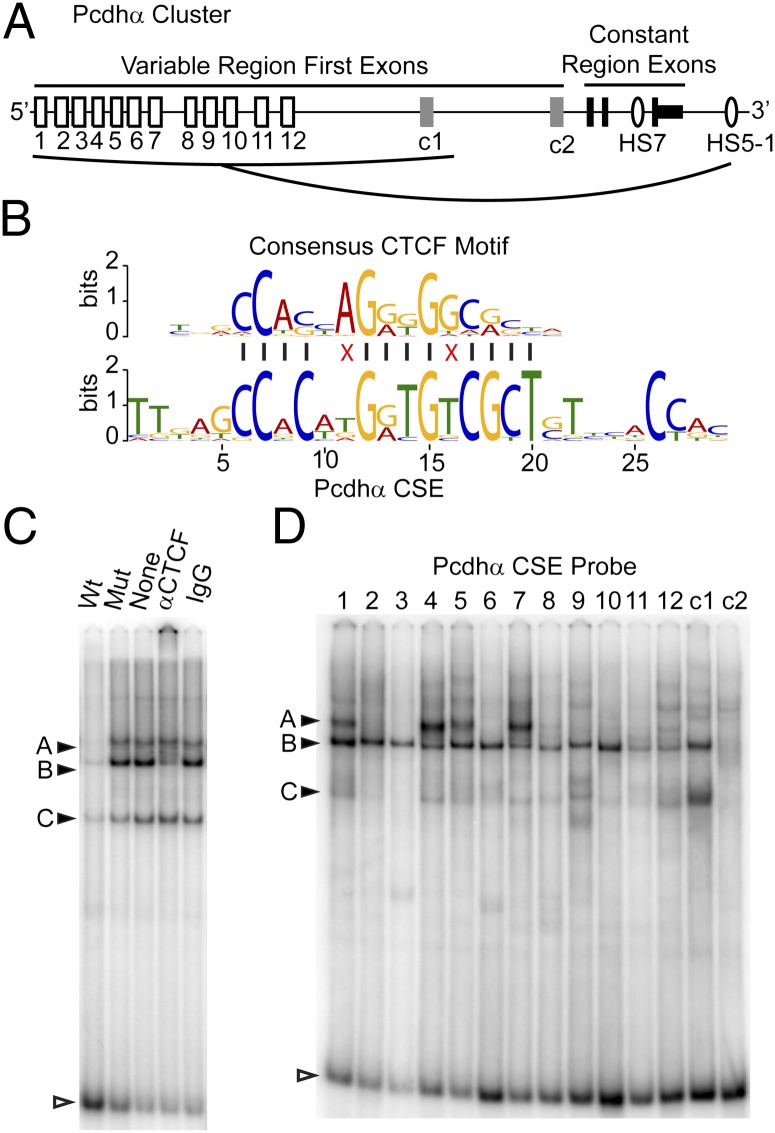
CTCF binds to the Pcdhα CSE in vitro. (A) The Pcdhα gene cluster showing the alternative “variable” first exons (white boxes), the c-type first exons (gray boxes), and the constant exons (black boxes). The variable exons regulated by HS5-1 are indicated by the line from HS5-1. (B) Alignment of the Jaspar core (30) CTCF motif with the Pcdhα CSE. Red “X’s” indicate mismatches. (C) EMSA using a α4 CSE probe with CAD nuclear extracts with different competitors: unlabeled wild-type (WT) or mutated (mut) α4 CSE, antibody to CTCF, or normal rabbit IgG. Filled arrowheads indicate protein-DNA bands and open arrowhead indicates free probe. (D) EMSA, as above, using radiolabeled probes encoding each Pcdhα CSE.

