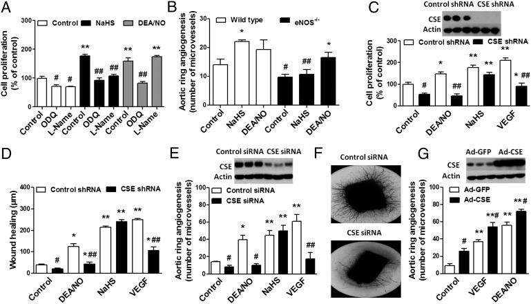
Fig. 1.
Mutual requirement of NO and H2S for angiogenesis in vitro. (A) bEnd3 cells were seeded overnight in 12-well plates, pretreated with ODQ (10 μM, 2 h) or l-NAME (4 mM, 40 min), and subsequently incubated with NaHS (30 μM) or DEA/NO (10 μM) for an additional 48 h. Cells were trypsinized and counted using a Neubauer hemocytometer. **P < 0.01 vs. control; ##P < 0.01 vs. NaHS. (B) Aortic rings were harvested from wild-type or eNOS−/− mice, cultured for 7 d in collagen gel in Opti-MEM medium containing 1% FBS, in the presence or absence of NaHS (30 μM) or DEA/NO (10 μM). *P < 0.05 vs. corresponding control; #P < 0.05 and ##P < 0.01 vs. corresponding wild type. (C) The lentiviral shRNA vector targeting CSE was transfected into bEnd3 cells. The shRNA vector effectively inhibited the expression of CSE gene in bEnd3 cell line at the protein level, as shown by Western blot analysis (C Upper). Following CSE silencing, cells were seeded in 12-well cell culture plates, and cell proliferation (C) and wound healing (D) were then evaluated in the presence of vehicle, DEA/NO (10 μM), NaHS (30 μM), or VEGF (20 ng/mL). *P < 0.05 and **P < 0.01 vs. control; #P < 0.05 and ##P < 0.01 vs. corresponding control shRNA. (E) Rat aortic rings were silenced with CSE siRNA for 48 h. Following gene silencing, aortic explants were placed individually on the bottom of 24-well plates, and collagen gel was gently applied. Rings were cultured for 7 d in the presence of vehicle, DEA/NO (10 μM), NaHS (30 μM), or VEGF (20 ng/mL). Western blots confirm efficient CSE silencing in aortic rings. CSE silencing markedly reduced both VEGF- and DEA/NO-induced vessel sprouting. *P < 0.05 and **P < 0.01 vs. control; #P < 0.05 and ##P < 0.01 vs. corresponding control siRNA. (F) Representative images of the 7-d collagen gel cultures of aortic rings exposed to VEGF following CSE gene silencing. (G) Aortic rings were exposed to adenovirus expressing GFP or CSE before embedding in collagen gel. Overexpression of CSE enhanced VEGF- or DEA/NO-induced neovessel growth. **P < 0.01 vs. control; #P < 0.05 vs. corresponding Ad-GFP. (Upper) Representative Western blot for CSE protein in rings exposed to adenovirus expressing GFP or CSE.

