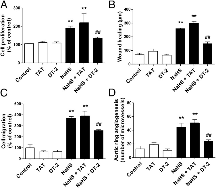
Fig. 3.
PKG-I inhibition prevents H2S-induced in vitro angiogenesis. (A) bEnd3 cells were pretreated with vehicle, DT-2 peptide (1 μM, 20 min), or the control peptide (TAT) (1 μM, 20 min) and then stimulated with NaHS (30 μM) to induce cell proliferation. The effect of DT-2 on NaHS-induced response was also estimated in wound healing (B) and cell migration (C). **P < 0.01 vs. control; ##P < 0.01 vs. NaHS + TAT. (D) Aortic ring explants were embedded in collagen gel, treated with the control peptide (TAT) or DT-2 (1 μM), and cultured for 7 d in the presence or absence of NaHS (30 μM, applied every 8 h). P < 0.01 vs. control; ##P < 0.01 vs. NaHS + TAT.

