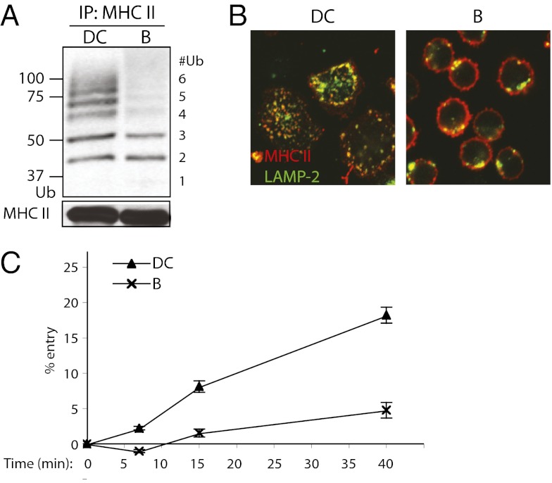
Fig. 1.
Differences in Ub chain length on MHC II of splenic DCs and splenic B cells correlate with differences in MHC II endocytosis. (A) Western blot of splenic DC and splenic B-cell (B) MHC II immunoprecipitates. MHC II was immunoprecipitated (IP) with TIB120 antibody; IPs were immunoblotted (IB) with anti-Ub antibody P4D1 and MHC II β-chain antibody Thorax. (B) Confocal microscopy of wild-type splenic DCs and splenic B cells. Cells were bound to coverslips, fixed in paraformaldehyde (PFA) and labeled with anti-MHC II antibody TIB120 (red) and lysosomal marker LAMP-2 (green). (C) Endocytosis of surface MHC II in splenic DCs and B cells. Cells were bound to anti-MHC II antibody TIB120 on ice and washed. This was followed by incubating the cells at 37 °C for the various times indicated to allow internalization of antibody-bound surface MHC II. An acidic wash (pH 3.0) was then used to strip the surface of remaining antibody. Internalized MHC II was protected from the acid stripping and detected by flow cytometry. Mean fluorescence intensity (MFI) values of MHC II and SEM were determined, and internalization is expressed as percentages of control MFI levels. Control cells were treated identically except for substituting a PBS wash for the acidic wash.

