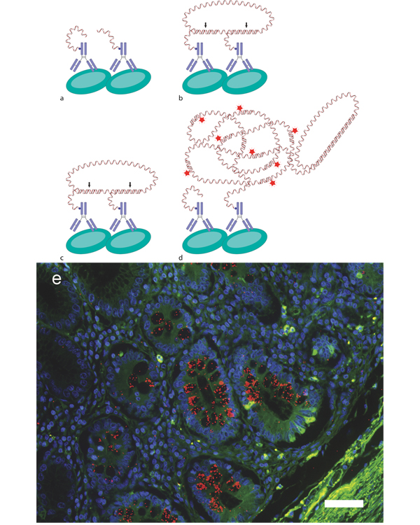
Figure 2.
Detection of protein interactions with in situ PLA. a Proximal binding of PLA-probes to interacting proteins will guide the b hybridization of circularization probes, c allowing them to be connected by ligation (arrows) and thereby creating a circular reporter molecule of the protein interaction. d The oligonucleotide on one of the PLA-probes will then act as primer for RCA to generate an RCA product that will be an elongation of the PLA-probe. Fluorescence-labeled detection oligonucleotides are then used to visualize the RCA-product. e Detection of Mucin2 glycosylation (Sialyl-Tn) (red dots) in formalin-fixed paraffin-embedded tissue sections from intestinal metaplasia [49]. Nuclei are counterstained with Hoechst 33342 (blue), using the autofluorescence of the tissue (green) to visualize the histology, scale bar equals 50 μm.

