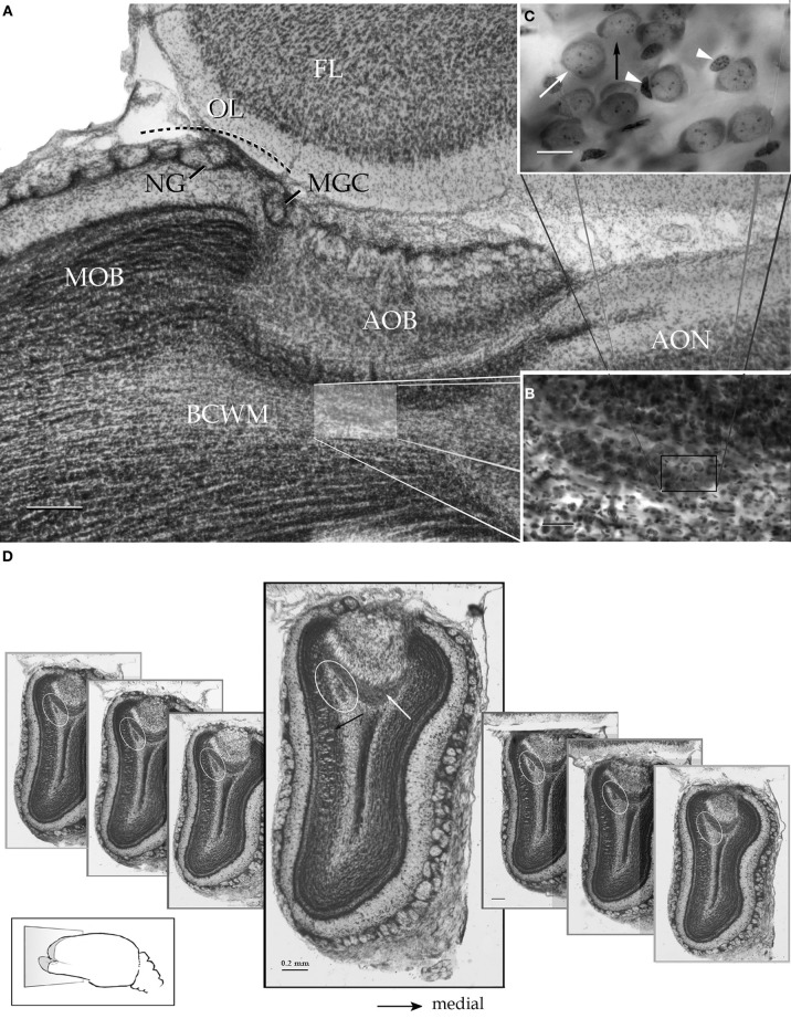
Figure 2.
Survey pictures of the olfactory bulb and the alpha component of the anterior olfactory nucleus as seen after thionine staining. (A) Sagittal section. The alpha component of the anterior olfactory nucleus (shaded) is embedded in the bulbar core white matter (BCWM), coursing horizontally, beneath the accessory olfactory bulb (AOB). FL, frontal lobe. (B) The spindle-shape alpha nucleus surrounded by the deep bulbar white matter is composed of dense packaged neurons. (C) High magnification micrograph from the alpha nucleus that is composed of polygonal (white arrow) and oblong, somewhat smaller, neurons (black arrow). The former neurons may exhibit an associated satellite cell (arrowheads). (D) Serial transverse sections from caudal (right) to rostral (left) showing the alpha component (encircled) in its rostrocaudal extent; note that the nucleus lies in a triangular area bounded by the granule cell (black arrow) and internal cellular (white arrow) layers of the main and accessory olfactory bulbs, respectively. Scale bars = 200 μm in A and D, 50 in B, and 10 in C. Nissl technique, adult rat brain.

