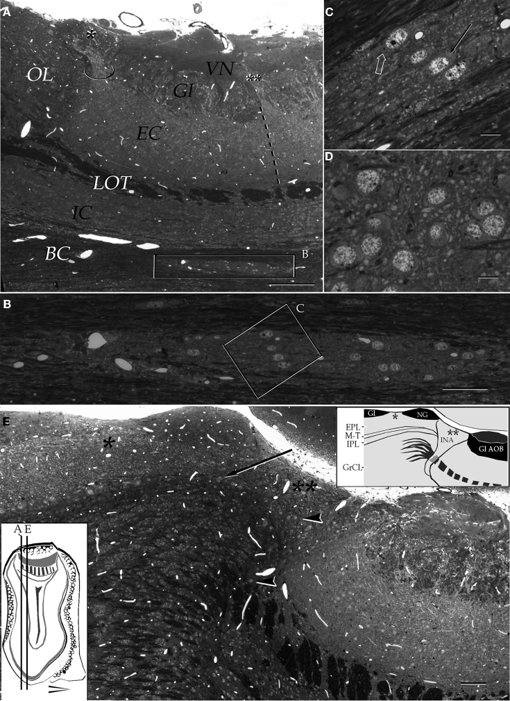
Figure 3.
Light micrographs from plastic-embedded longitudinal sections through the olfactory bulb. (A) Section at the caudal intersection of the main olfactory bulb with the accessory olfactory bulb (right side). Note the dendritic bundle (circle) arising from the external cellular layer (EC) and resolving at the base of a modified glomerulus (asterisk), adjacent to the olfactory limbus (OL). Beneath, embedded in the bulbar core white matter (BC), lies the alpha component of the anterior olfactory nucleus (boxed). VN, vomeronasal nerve layer; Gl, glomerular layer; LOT, lateral olfactory tract; IC, internal cellular layer. At the glomerular-free area of the accessory olfactory bulb the linea alba (dashed) separates the external cellular layer in a rostral (left) and caudal halves. (B) Higher magnification of the alpha component of the anterior olfactory nucleus. (C) Neuron somata from the alpha component of the anterior olfactory nucleus (boxed in B). A neuronal cluster made up of medium-sized neurons (solid arrow) and a large neuron whose cytoplasm contains Nissl substance (hollow arrow). (D) Neuron somata in the dorsal part of the anterior olfactory nucleus at the same magnification as the micrograph shown in C; note the difference in size and distribution of neurons in comparison to those from the alpha component shown in C. (E) Micrograph depicting the olfactory limbus between the main (left) and accessory olfactory bulbs. At the upper left is the caudal end of the main olfactory bulb, followed by the glomerular-free preolfactory (asterisk) area. The latter faces a second area or the necklace glomeruli whose external plexiform (EPL) and mitral/tufted cell (M-T) layers become indistinct and hypocellular (arrow), respectively. Following caudally is a wedge-shaped or interstitial area (double asterisik) (INA) that lacks laminar organization, having clusters of cells of assorted size (arrow-heads). Gl-AOB, glomerular layer of the accessory olfactory bulb; IPL, internal plexiform layer; GrCL, granule cell layer. Scale bars: 100 μm in A, 40 in B and E, and 10 in C and D. One-micrometer-thick sections, adult rat brain, toluidine blue staining.

