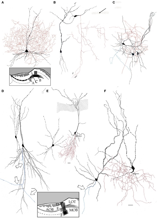
Figure 8.
High magnification drawings from bulbar neurons with projecting (blue) and local (red) axons. Somata and dendrites from the latter are nearly identical in shape, thickness, and varicosities. (A) Main accessory cell of the accessory olfactory bulb with a profuse axonal (red) field. (B) Deep short axon cell whose descending axon (red) distributes in the granule cell layer of the main olfactory bulb. (C) Two interstitial bulbar interneurons with ascending axon and a projecting cell whose axon (green) divides into two descending collaterals. (D) Examples of interstitial neurons. The projecting cell (right) emits a descending axon (arrows). (E) A projecting cell whose parent axon provides a huge number of collaterals to the adjacent neuropil. (F) Pyramidal-like and a short-axon neurons in the alpha component of the anterior olfactory nucleus. Adult rat brain rapid-Golgi method. Scale bar = 15 μm.

