Figure 3.
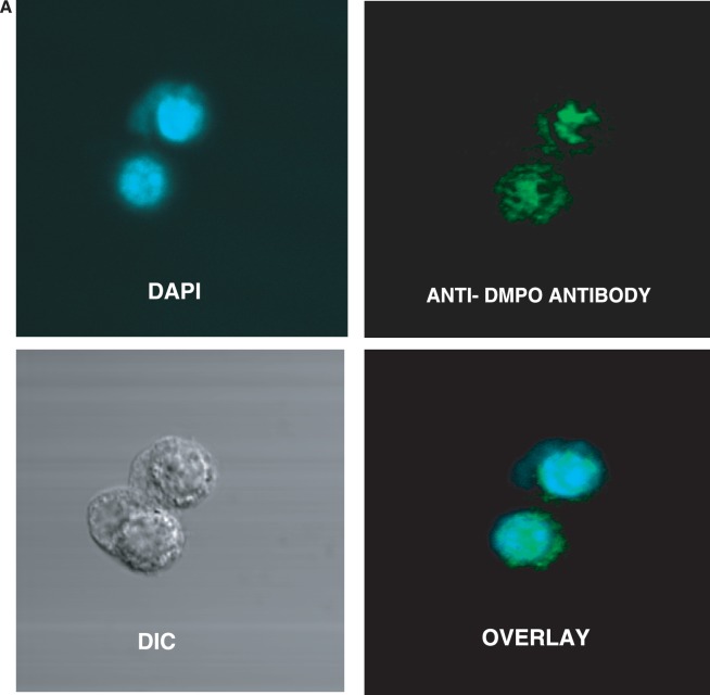
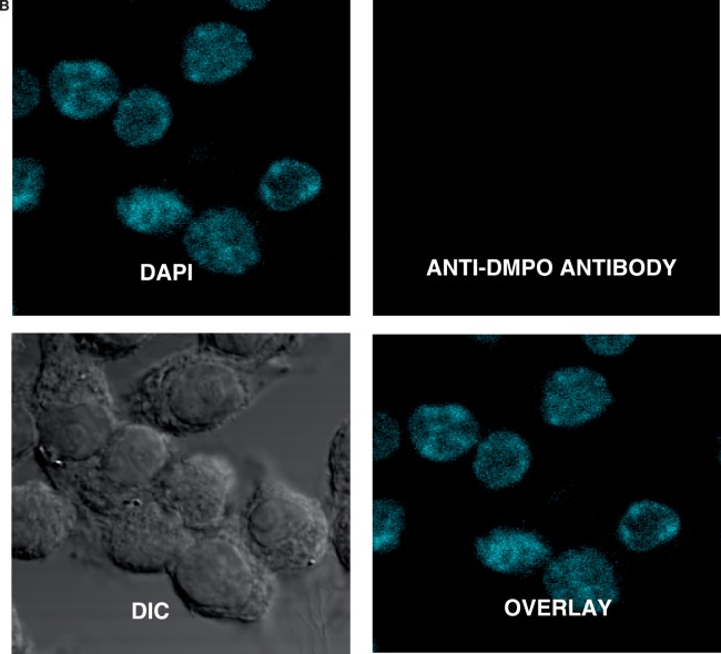
Confocal laser scanning microscopy of DNA-derived radical adducts in RAW 264.7 cells. Adherent RAW 264.7 cells in glass-bottomed 32 mm2 dishes were treated with CuCl2/H2O2 in the presence of 25 mM DMPO for 1 h. Cells were fixed with paraformaldehyde and stained with the anti-DMPO antibody, which was detected with an Alexa 488 conjugated secondary antibody. (A) Cells were imaged and anti-DMPO immunoreactivity (green stain) could be seen primarily in the nucleus (DAPI, blue stain), as evidenced by the co-localization (overlay image). (B) There was no DMPO immunoreactivity in cells that were not treated with the spin trap.


