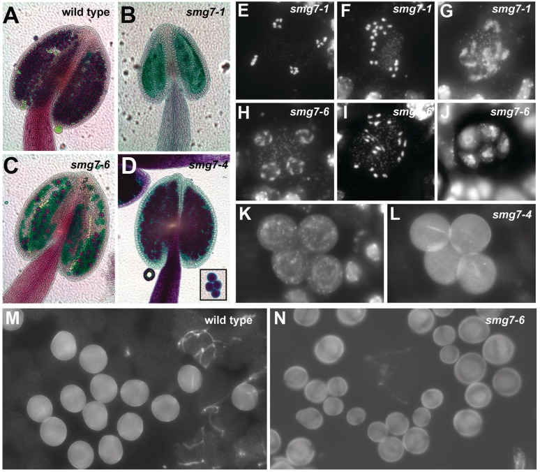
Figure 2.
Development of male germ-line in smg7-6 and smg7-4 mutants. (A–D) Anthers of different smg7 mutants after Alexander staining. Viable pollen stain red, no pollen is present in smg7-1 anthers. Inset in (D) shows four joint pollen grains in smg7-4 mutants. (E–J) End of second meiotic division in smg7-1 and smg7-6 mutants. Meiotic chromosomes are stained with DAPI: (E) regular anaphase II, (F, I) irregular anaphase II, (G) slowly decondensing chromosomes after anaphase II, (H) regular telophase II, (J) a polyad consisting of five separated cells with irregular nuclei. (K, L) Microspores typical for smg7-4 mutants remain attached after meiotic division. Nuclei are stained with DAPI (K), cell walls detected due to their auto-fluorescence using FITC filters (L). (M, N) Fields of microspores in wild-type and smg7-6 mutants.

