Figure 2.
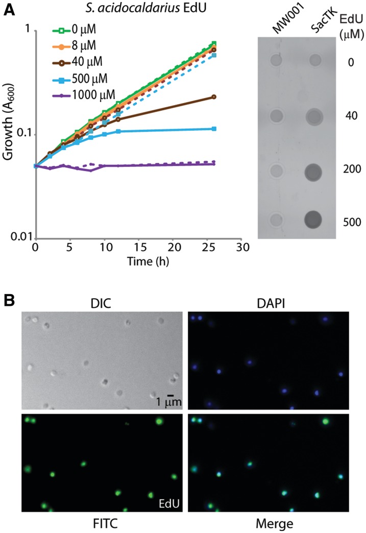
Sulfolobus acidocaldarius cells expressing FLAG-TK incorporate EdU into genomic DNA. (A) MW001 (dashed lines) and SacTK (solid lines) cells were grown in medium containing the indicated concentrations of EdU and growth was monitored by A600. Genomic DNA was extracted from 24 h samples and digested with PstI. Incorporated EdU was fluorescently labelled using ‘click’ chemistry. Three hundred nanograms of each DNA sample were spotted onto a nitrocellulose membrane and visualized using a FLA5000 Phosphoimager. (B) Fluorescent microscopy of SacTK cells grown for 3 h with 100 µM EdU. After fixing with 2.5% paraformaldehyde, cellular DNA was fluorescently labelled using ‘click’ chemistry. Images show DIC, DAPI staining of DNA (blue), EdU-AlexaFluor488 (green) and merged images.

