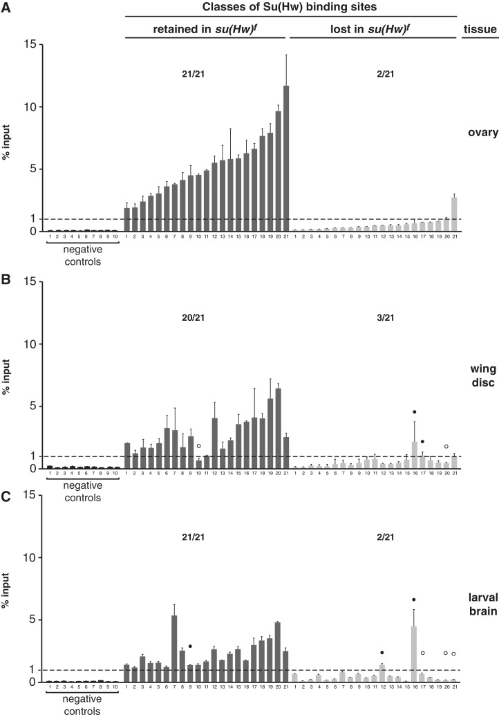
Figure 7.
Su(Hw)f binding is retained at tissue-specific sites. (A–C) ChIP-qPCR analyses of Su(Hw)f binding to the same set of 21 sites in chromatin isolated from the ovary (A), third instar wing disc (B) and third instar larval brain with eye and antennal disc (C). Negative controls represent sites that lack a Su(Hw)-binding motif and were not identified in the ChIP-Seq dataset. The dashed line indicates 1% input threshold. SBSs are divided into f-retained (dark gray, 21 sites) and f-lost (light gray, 21 sites). Averaged values of two biological replicates are shown, error bars indicate standard deviation. For each experiment, the ratio represents the number of SBS above 1% input level over the total number of sites tested. White and black circles indicate tissue-specific f-lost and f-retained sites, respectively.

