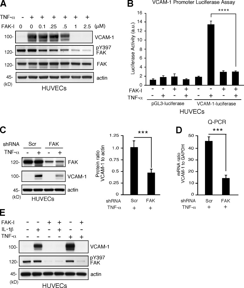
Figure 3.
FAK activity is essential for proinflammatory cytokine-mediated VCAM-1 expression. (A) TNF-α–induced (10 ng/ml, 16 h) VCAM-1 is prevented by FAK-I (PF271) treatment of HUVECs in a dose-dependent (0.1–2.5 µM FAK-I) manner. VCAM-1, pY397 FAK, total FAK, and actin levels were determined by immunoblotting. (B) TNF-α–induced VCAM-1 promoter activity is blocked by FAK inhibition. HUVECs were transfected with pGL3 promoterless luciferase (control) or pGL3 fused with the human VCAM-1 promoter, stimulated with TNF-α, and treated with FAK-I (1 µM PF271), as indicated, and relative luciferase activity (arbitrary units [a.u.]) is shown. Values are means (±SD) from three independent experiments. (C) FAK knockdown prevents VCAM-1 expression. HUVECs were transduced with lentiviral Scr or anti-FAK short hairpin RNA (shRNA) and stimulated with TNF-α. FAK and VCAM-1 and actin expression levels were determined by immunoblotting, and densitometry values of VCAM-1 relative to actin are means (±SD; n = 3; ***, P < 0.001). (D) VCAM-1 mRNA levels to GAPDH were determined by Q-PCR (±SD; n = 3; ***, P < 0.001) in experiments, as described in C. (E) FAK activity is required for IL-1β–induced VCAM-1 expression. HUVECs were stimulated with 20 ng/ml IL-1β and 10 ng/ml TNF-α, and FAK-I (1 µM PF271) was added as indicated. After 16 h, VCAM-1, pY397 FAK, and actin levels were evaluated by immunoblotting.

