Figure 5.
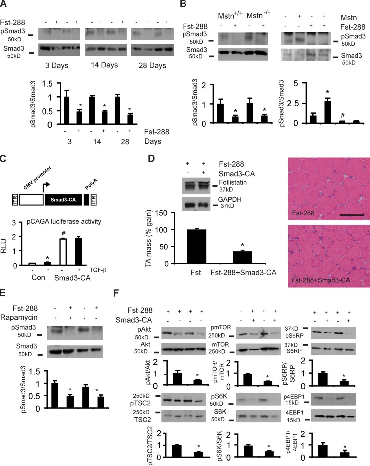
Smad3 regulates Fst-induced hypertrophy and is a key intermediary that regulates PI3K/mTOR signaling. (A) Smad3S423/425 phosphorylation is reduced at 3, 14, and 28 d after Fst-288 administration (*, P < 0.05 vs. control muscles). (B) Smad3 phosphorylation is reduced 28 d after rAAV6:Fst-288 administration to the muscles of myostatin-null mice (Mstn−/−), similarly compared with the treated muscles of littermate controls (Mstn+/+; *, P < 0.05 vs. control muscles). Injection of the muscles of wild-type mice with an rAAV6 vector expressing myostatin (Mstn) increases Smad3 phosphorylation, but coinjection of muscles with rAAV6:Mstn and rAAV6:Fst-288 still results in diminished Smad3 phosphorylation (shown here after 28 d; *, P < 0.05 vs. control muscles; #, P < 0.05 vs. control muscles). (C) Smad3-CA was validated in 293T cells using a pCAGA luciferase assay normalized to β-gal expression. (D) 8-wk-old mice were injected with either rAAV6:Fst-288, or rAAV6:Fst-288 and rAAV6:Smad3-CA, and examined after 28 d (*, P < 0.001 vs. control). Data are expressed as the percentage increase in muscle mass relative to the control leg, where the effect of Fst-288 is normalized to 100%. Hematoxylin and eosin–stained cryosections demonstrate attenuated muscle fiber hypertrophy in muscles examined 28 d after receiving rAAV6:Fst-288 and rAAV6:Smad3-CA compared with rAAV6:Fst-288 alone. Bar, 100 µm. (E) Smad3 phosphorylation in response to rAAV6:Fst-288 injection was assessed in mice additionally treated with vehicle or rapamycin for 14 d (*, P < 0.05 vs. control muscles). (F) Akt/mTOR signaling was assessed 28 d after the co-administration of Fst with Smad3-CA (*, P < 0.01 vs. control muscles). Graphs show data from at least four independent experiments. Error bars indicate ± SEM.

