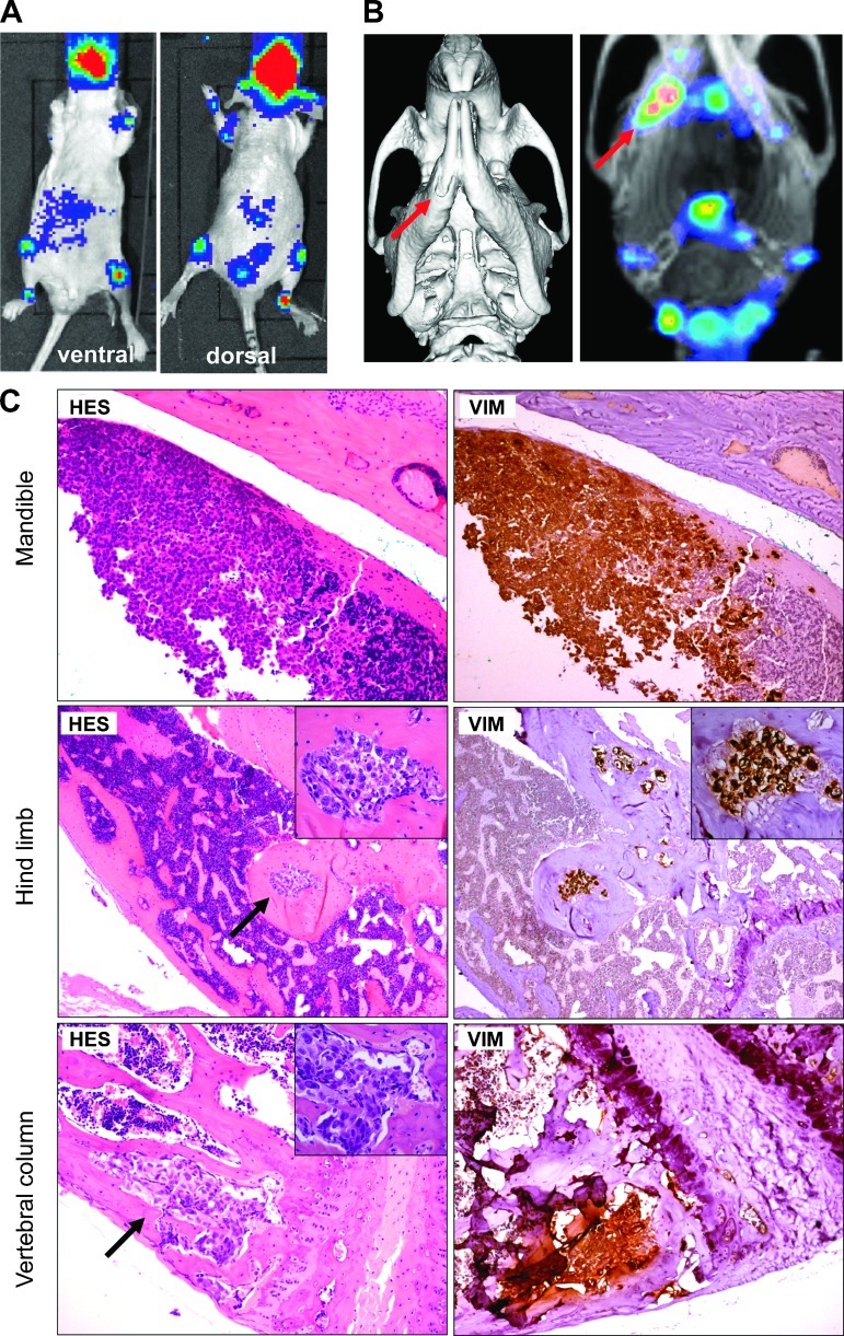
Figure 5.
IGR-CaP1 cells generate bone metastases after intracardiac injections. IGR-CaP1 cells were injected into the left cardiac ventricle. (A) Multiple bone metastases were observed using BLI 5 weeks after injection. A representative mouse showed the bone localization of metastases. (B) CT scan acquisition and the incorporation of 99mTc-MDP measured with SPECT confirmed the presence of bone metastases in the mandible. (C) Histologic staining of decalcified bone sections confirmed the presence of bone lesions. As shown with intratibial injection, metastases in limb were osteoblastic. All bone metastases showed intense VIM expression. Magnification, x50; insert, x400. Arrows show the metastases.

