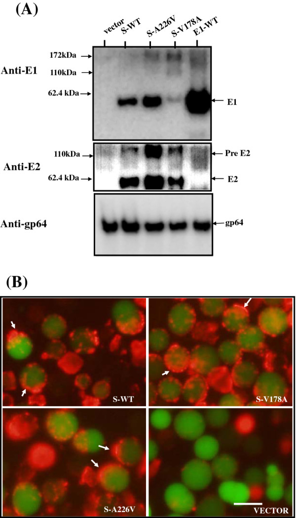
Figure 5.
E1 co-expression with other structural proteins by 26 S-based constructs. Sf21 cells were grown in a six-well plate and infected with recombinant viruses at an M.O.I. of one. E1 and E2 proteins were detected by Western blot analysis using rabbit anti-CHIKV E1 serum (upper gel), or anti-CHIKV E2 serum (middle gel) then re-probing with anti-baculovirus gp64 antibody (lower gel). Proteins extracted from cells were infected with various baculoviruses as indicated above the gel. Arrows on the right indicate the CHIKV E1 and E2 proteins. Two protein size markers are shown on the left. (B) Immunofluorescence analysis of CHIKV E1 on the cell surface. Sf21 cells infected with the indicated recombinant baculoviruses and described as above were stained as per Figure 3C. The bar represents 10 μm.

