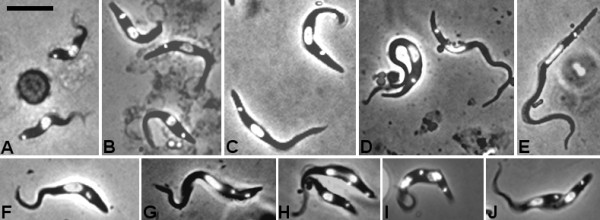
Figure 1.

Morphology of developmental stages ofTrypanosoma congolense. Fixed and DAPI-stained cells; each panel shows a merge of DAPI and brightfield images. A. Bloodstream forms. Panels B to F are procyclic trypomastigotes from the tsetse midgut 2 days (B), 6 days (C), 9 days (D) and 17 days (E) after ingestion of the infected bloodmeal. Panels F to J are trypomastigotes from the tsetse midgut in various stages of division; 2K1N (F, G), 2K2N (H-J). Bar = 10 μm.
