
Figure 5.
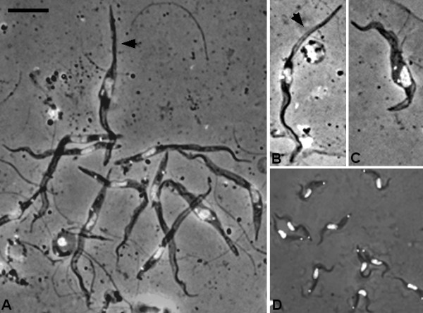
Trypanosomes in spit samples 16–21 days after infection. A. Mixture of trypomastigotes with epimastigote (arrowed). B. Epimastigote with long posterior (arrowed). C. Epimastigote (2K1N). D. Metacyclics from day 21; contrast the size of metacyclics with the other trypomastigotes from the proboscis shown in panel A at the same scale. Bar = 10 μm.