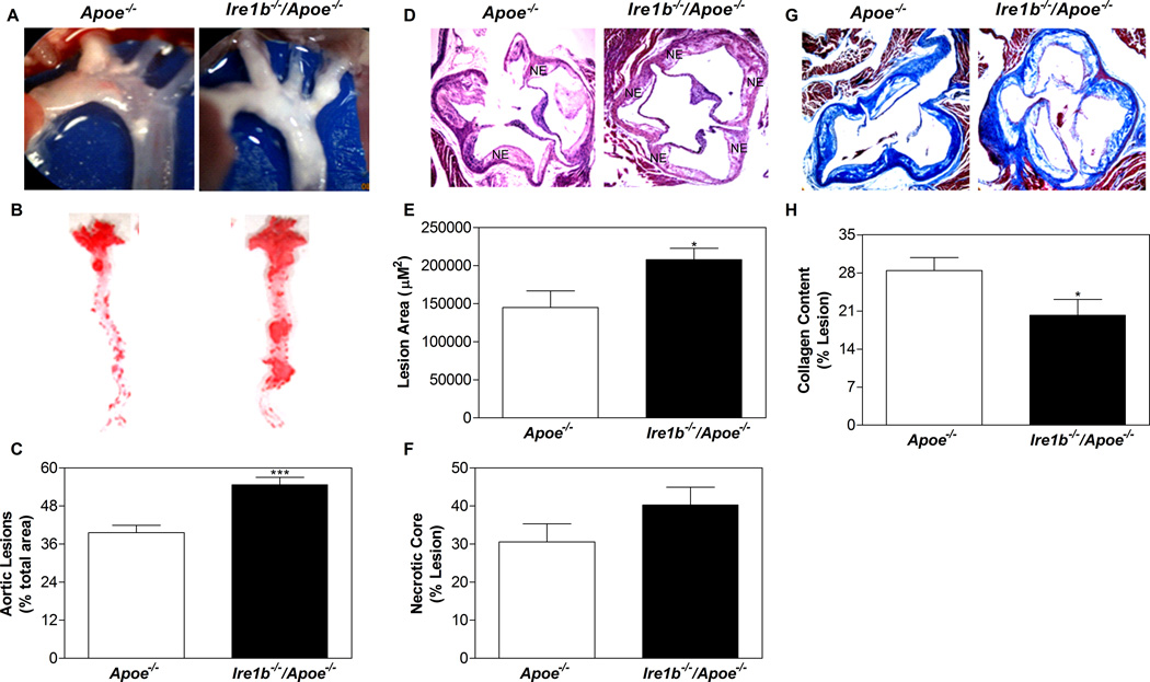
Figure 3. Ablation of IRE1β in Apoe−/− mice enhances atherosclerosis.
Ire1b−/−/Apoe−/− and Apoe−/− mice were fed a chow diet (n=8) for 12 months. (A) Aortic arch and other proximal arteries were dissected and photographed. A representative photograph from each group is shown. (B–C) Aortas were isolated, stained with Oil Red O (B) and quantified (C). (D–F) Hematoxylin and eosin staining (D) of the proximal aorta (root assay) showing necrotic core (NE) is shown. Quantitative analyses of the lesion (E) and necrotic core (F) were done by Image-Pro-Plus 4.5 software. (G–H) Collagen was stained with Masson’s trichrome (G) and quantified using Image J (H). Values are mean ± SD. *p<0.05, **p<0.01, and ***p<0.001 compared to Apoe−/−.

