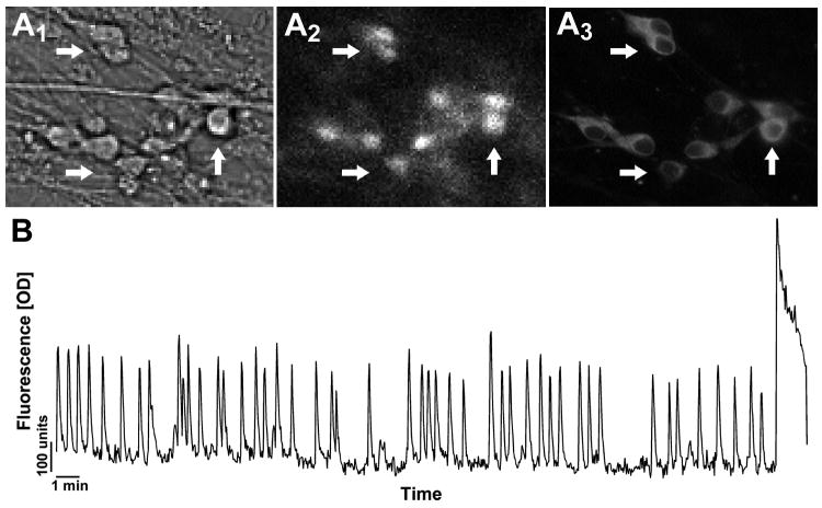
Figure 4.
Calcium Green-1 imaging of GnRH neurons 6-10 days in vitro. A Cells were identified by their bipolar morphology (A1), loaded with fluorescent calcium-sensitive dye (A2), and their identity verified post-imaging by immunocytochemistry (A3). Arrows indicate identical cells in all fields. B Representative recording showing spontaneous baseline oscillations in intracellular calcium levels in a GnRH neuron during 33-min in serum free media (Y-scale = OD units).

