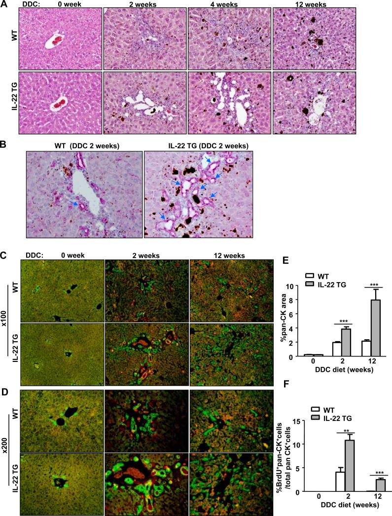
Fig. 2. The IL-22TG mice have increased LPC proliferation after being fed a DDC diet compared with the WT mice.
The mice were fed a DDC diet for up to 12 weeks. (A) H&E staining of liver tissues from DDC-fed mice at different time periods. (B) Representative immunohistochemical analyses with an anti-pan-CK antibody of liver tissues from mice fed a DDC diet for 2 weeks. The arrows indicate ductular reactions. (C, D) Pan-CK (green)/BrdU (red) double staining of liver tissues from mice fed a DDC diet for 2 or 12 weeks with a BrdU injection 2h before sacrifice. (E, F) The number of pan-CK+ and BrdU+pan-CK+ double positive cells was quantified. **P<0.01 and ***P<0.001. Representative photographs from three independent experiments with similar results are shown.

