Figure 1.
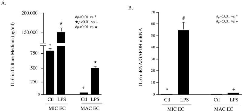
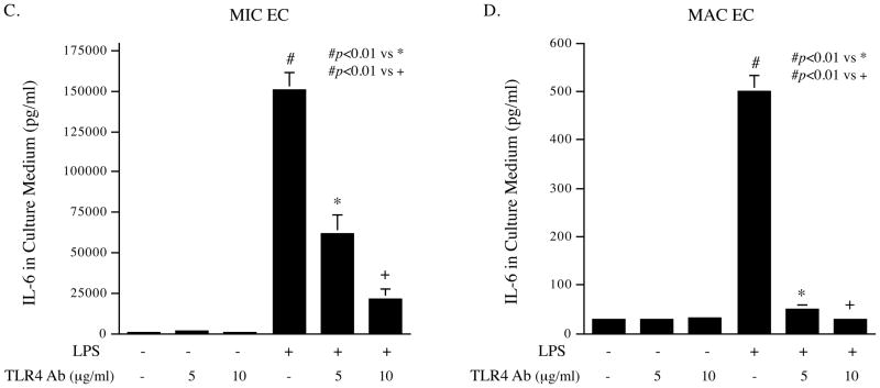
Stimulation of IL-6 expression by TLR4 activation in MIC and MAC ECs. A and B: 100% confluent cultures of MIC or MAC ECs were treated with or without 100 ng/ml of LPS for 24 h. After the treatment, IL-6 in culture medium was quantified with ELISA (A) and IL-6 mRNA in cells was quantified with real-time PCR (B). C and D: MIC ECs (C) or MAC ECs (D) were treated with or without 100 ng/ml of LPS in the absence or presence of 5 or 10 μg/ml of anti-TLR4 antibody (Ab) for 24 h. After the treatment, IL-6 in culture medium was quantified with ELISA. The presented data were from representative of three experiments with similar results.


