
Fig. 2. GFP-expressing neuralized mouse embryonic stem cells (nESCs) co-localize with RFP-expressing human tumor cells in an organotypic brain slice.
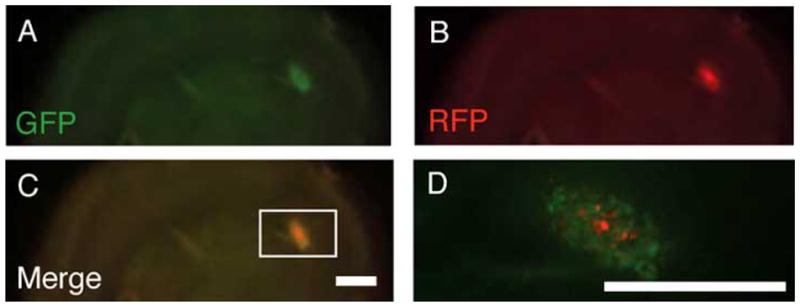
Stereoscopic epifluorescent images show tumor mass infiltration by nESCs. GFP-expressing nESCs and RFP-expressing human glioma cells were introduced on the surface of the organotypic rat brain slice. The living brain section was obtained from a 300-micron thick coronal slice just anterior to bregma obtained from a fresh rat brain embedded in agarose and cut with a vibratome. Initially, 10 μl (~8,000 cells/μl) of both cell types were simultaneously implanted at separate locations on the surface of a one-week old slice. Aliquots of the two cell types were applied approximately 10 mm from each other across the width of the organotypic slice. Images shown here were taken one week after implantation of the nESCs and glioma cells. (A–D) Stereomicroscopic epifluoresent images show that stem cells placed distant from the tumor cells co-localized with tumor cells and were not detectable in other regions of the brain slice. Scale bars = 1 mm; Scale bar in C applies to A–C.