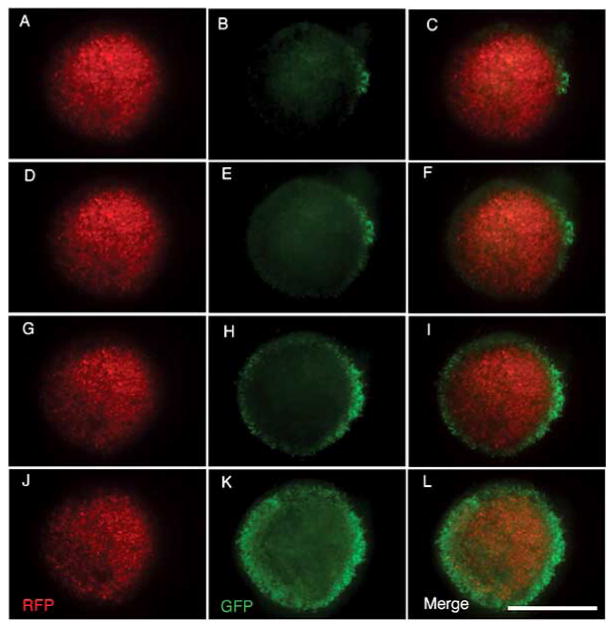
Fig. 3. Time course of tumor infiltration by neuralized embryonic stem cells on an organotypic brain slice.
These stereoscopic epifluorescent images show the progression of tumor mass infiltration by nESCs over the course of a several weeks in an organotypic brain slice culture. GFP-expressing nESCs and RFP-expressing human glioma cells were introduced on the surface of an organotypic rat brain slice as described in Fig. 2. (A–C) Starting at 2 weeks post-implantation, nESCs are found at the tumor mass. At 3 weeks (D–F) and 4 weeks (G–I) post-implantation, the tumor mass becomes infiltrated progressively by nESCs. At 6 weeks (J–L), the tumor mass becomes encapsulated completely by the stem cells, suggesting contiguity between nESCs and glioma cells. Scale bar = 1 mm; Scale bar applies to all panels.

