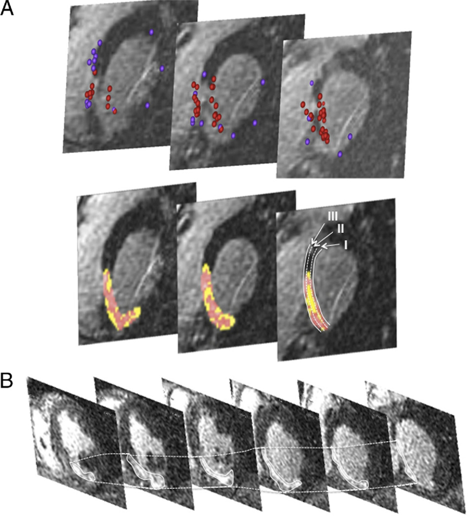
Figure 5. Stack of Short-Axis Delayed-Enhanced Magnetic Resonance Images.
(A) Magnetic resonance imaging with delayed enhancement (stack of short-axis views from a basal [right] to an apical direction) in a patient with intramural ventricular tachycardia. This patient had an inferoseptal myocardial infarction. The scar extends from the basal septum to the midinferior septum. Mapping points (upper panel) obtained during electroanatomic mapping were projected onto the magnetic resonance imaging (red tags indicate low-voltage sites, purple tags indicate normal voltage sites) confirming the presence of endocardial scar tissue. The lower panel shows the automated color coding depending on the signal intensity. The septum was divided into an endocardial third (I), an intramural third (II), and an epicardial third (III). The scar was also separated into core infarct (red) and peri-infarct zone (yellow). (B) Delayed-enhancement magnetic resonance imaging (stack of short-axis views from a basal [right] to an apical direction) in a patient with an inferoseptal infarction and endocardial ventricular tachycardia. No intramural ventricular tachycardia circuit was present in this patient. Compared with A, the intramural segment lacks areas with tissue heterogeneity.

