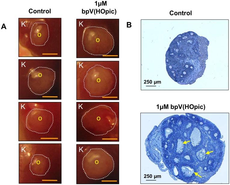
Figure 1. Enhanced follicular development by transient treatment of neonatal mouse ovaries with the PTEN inhibitor bpV(HOpic).
(A) Comparison between the sizes of treated and control ovaries transplanted under the kidney capsules. One ovary from a PD3 mouse was cultured for 24 h with 1 µM bpV(HOpic) and another ovary was cultured without bpV(HOpic) and then transplanted under the capsule of each kidney of the same ovariectomized recipient as described in Materials and Methods. Ovaries that were treated with bpV(HOpic) before transplantation grew bigger than the non-treated control ovaries. K represents kidney tissue from the recipient, O represents the transplanted ovary, and the ovarian border is outlined by dashed circles. Scale bar = 1 mm. (B) Morphological analysis of treated and control ovaries excised from the kidney capsules. Ovaries from PD3 mice were cultured for 24 h with or without 1 µM bpV(HOpic) before transplantation under each kidney capsule of the same ovariectomized recipient as described in Materials and Methods. One day after the transplantation, recipient mice were treated daily with 2 IU of pregnant mare serum gonadotropin for 18 days. Fourteen hours before being killed, the mice were treated with 5 IU of human chorionic gonadotropin. Ovaries were excised from the kidney capsules and embedded in paraffin, and serial sections of 8 µm thickness were prepared and stained with hematoxylin. A larger number of antral follicles were observed in the bpV(HOpic)-treated ovaries (arrows) than in the control ovaries. The experiments were repeated at least 4 times, and 5 mice were used each time. Scale bar = 250 µm.

