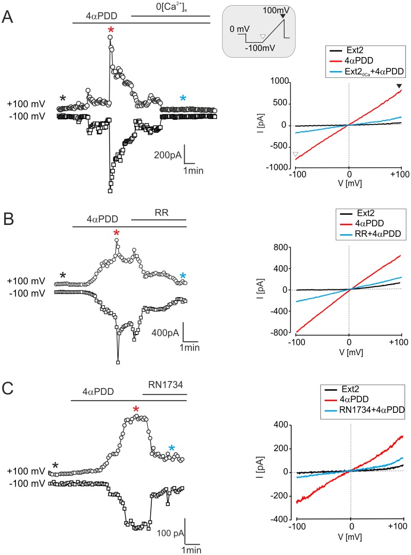
Figure 10. Currents evoked by 4αPDD in astrocytes in vitro are reduced by calcium-free extracellular solution, Ruthenium Red or RN1734.
(A–C, left) Time course of 4αPDD-evoked currents measured from the ramp protocol in astrocytes isolated from the hippocampus 7D after H/I (for the voltage protocol see the inset) prior to and during 4αPDD (5 µM) application and after removing extracellular Ca2+ (Ext2ØCa, A) or after the application of TRPV4 inhibitors, such as Ruthenium Red (RR, 10 µM, B) or RN1734 (10 µM, C). (A–C, right) The traces of steady state currents (same cells as in left) obtained in Ext2 solution (black lines), during 4αPDD application (red lines) and after removing extracellular Ca2+ (Ext2ØCa, A) or after the application of TRPV4 inhibitors, such as Ruthenium Red (RR, 10 µM, B) or RN1734 (10 µM, C), are indicated by blue lines. Representative traces of steady state currents were obtained at the times indicated by asterisks of the corresponding colors.

