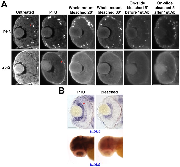
Figure 8. Performance of immunostaining and in situ hybridization after bleaching.
(A) Immunostaining experiments were conducted with untreated, PTU-treated or bleached embryos at 52 hpf. Two first antibodies, anti-PH3 and zpr2 were used. The typical positive signal is indicated by a red arrowhead in each case. Scale bar = 50 µm. (B) An in situ hybridization experiment with tubb5 using PTU-treated or bleached embryos. These samples were obtained from a double in situ hybridization experiment with another gene stained in red; thus the samples, especially the whole-mount embryos, look reddish in general. Scale bar = 50 & 100 µm for sectioned and whole-mount samples respectively. All images were acquired with the same parameters and were not altered during the figure composition to ensure comparability across conditions.

