
Figure 3. EMSA of wild type and mutant Czf1p binding to the CZF1 promoter region.
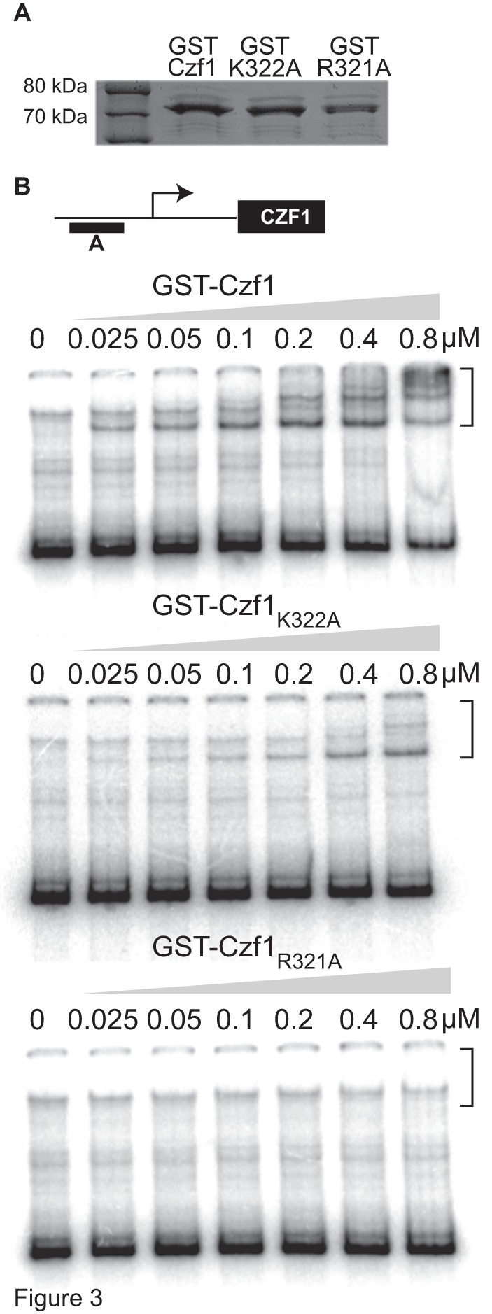
(Panel A) SDS PAGE analysis of 500 ng of purified wild type and mutant GST-Czf1 fusion proteins stained with Coomassie Blue. (Panel B) Diagram of the CZF1 promoter region (not drawn to scale). The DNA fragment used in this study (fragment E, 565 bp) is represented as a black rectangle and is located −3381 to −2816 from the ATG start of the CZF1 ORF. The transcriptional start site is represented as an arrow and is located at −2065 from the ATG [23]. (Panels C–E) Increasing amounts of GST-Czf1p were incubated with 32P end-labelled fragment E. The samples were analyzed by electrophoretic mobility shift assay and phosphorimaging. Brackets indicate shifted fragments. All EMSA experiments were repeated at least 3 times and a representative experiment is shown.