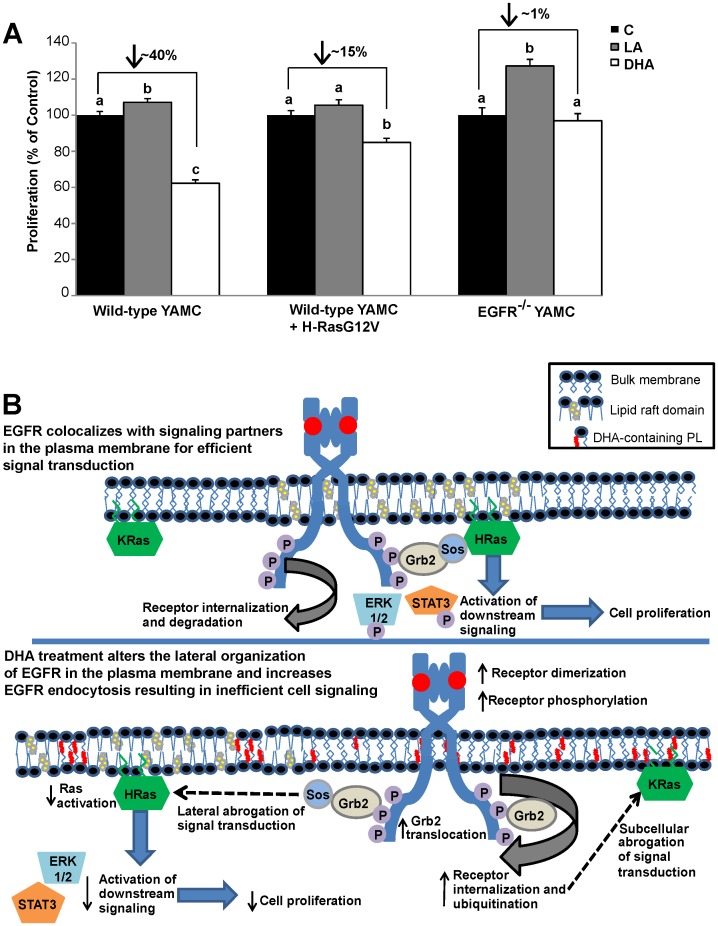
Figure 7. DHA suppresses EGFR-mediated cell proliferation.
A) Wild-type and EGFR−/− YAMC cells were treated with 50 µM BSA-complexed fatty acid for 24 h. Following the initial treatment, cells were seeded at an equal density into a 96-well plate, and cultured under the same treatment conditions for an additional 48 h. Select cultures were transfected using nucleofection with GFP-H-RasG12V prior to being seeded into the 96-well plate. Cell proliferation was measured using CyQUANT cell proliferation assay. Results are representative of 3 independent experiments. Data are expressed as mean±SEM (n = 9 samples per treatment). Statistical significance between treatments as indicated by different letters (P<0.05) was determined using ANOVA and Tukey’s test of contrast. No comparisons were made between proliferation of wild-type and EGFR−/− cells. B) Putative model: Under control conditions, EGFR is enriched in liquid ordered lipid raft domains of the cell. Upon stimulation, ligand bound receptors dimerize and transphosphorylate. The phosphorylated tyrosine residues then serve as docking sites for adaptor proteins, which leads to the activation of downstream targets, including Ras. Signaling is efficient due to the lateral organization of EGFR and downstream signaling partners. Upon fatty acid treatment, the cell incorporates DHA into plasma membrane phospholipids. DHA is sterically incompatible with cholesterol and perturbs liquid ordered domains resulting in a change in the lateral organization of EGFR. Ligand-induced receptor dimerization and phosphorylation is increased, along with receptor ubiquitination, internalization, and degradation. The altered lateral and subcellular organization of EGFR results in inefficient cell signaling. Suppression of EGFR signaling causes altered cellular function, including reduced cell proliferation.

