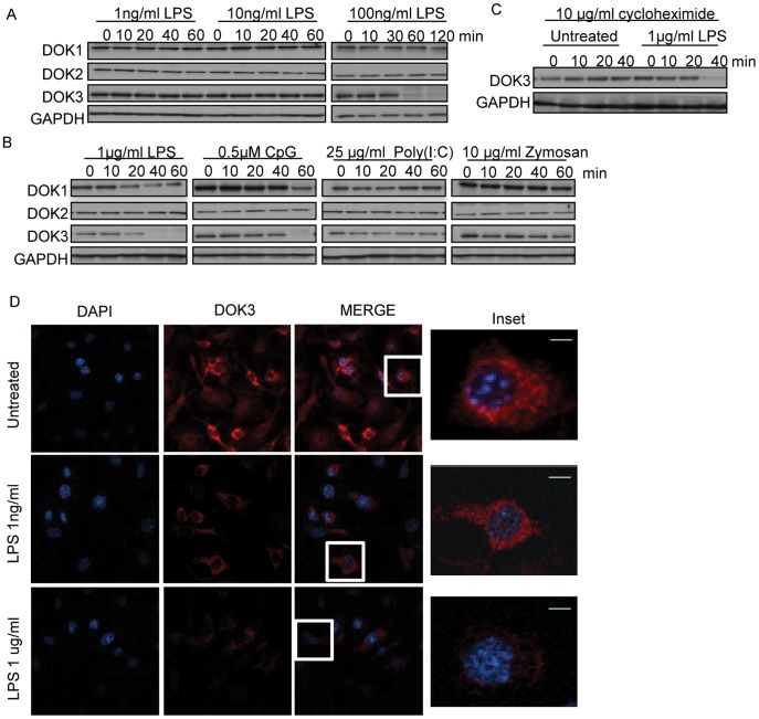
Figure 1. LPS stimulation of macrophages induces DOK3 degradation.
BMM were stimulated with (A) 1, 10, or 100 ng/ml LPS; or (B) 1 µg/ml LPS (TLR4 ligand), 0.5 µM CpG (TLR9 ligand), 25 µg/ml Poly (I:C) (TLR3 ligand), or 10 µg/ml Zymosan (TLR2 ligand) for the indicated time and cell lysates were immunoblotted for DOK1, DOK2, DOK3, and GAPDH. (C) BMM were treated with 10 µg/ml of the translational inhibitor cycloheximide during LPS stimulation. Data are representative of three independent experiments. (D) BMM cells were stimulated with LPS (1 ng/ml and 1 µg/ml) for 1 h. The cells were fixed, permeabilized, and immunofluorescent staining for DOK3 (red) and DAPI (blue) was performed and visualized with a Leica TCS NT Confocal microscope. Bar is 10 µm.

