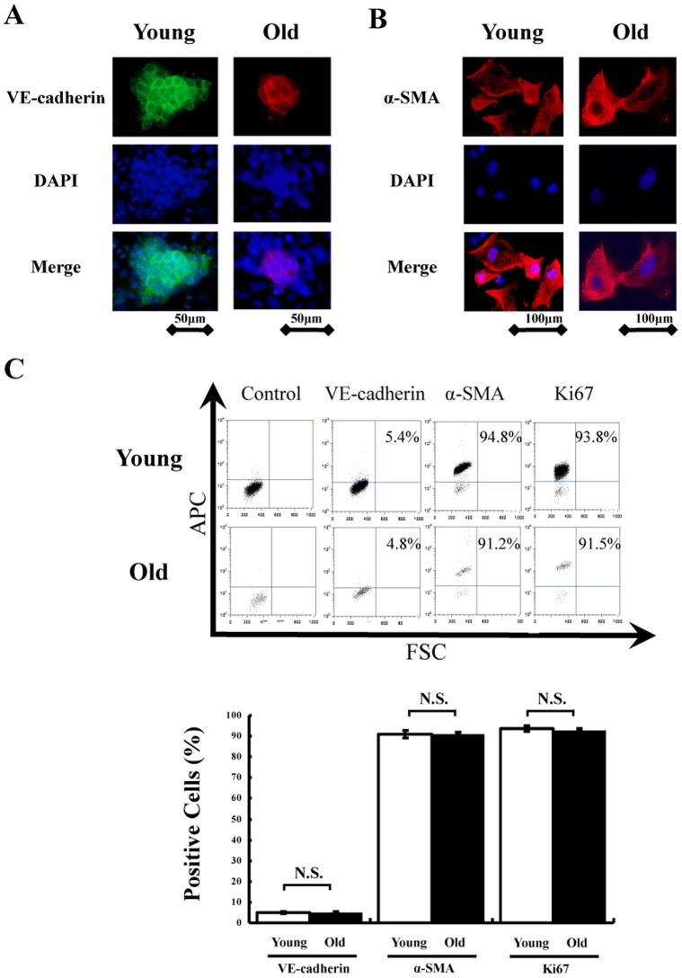
Figure 1. Differentiation into mature vascular cells in vitro.
Sorted Flk-1+ cells derived from young and old iPS cells successfully differentiated into (A) mature endothelial cells (VE-cadherin positive) and (B) smooth muscle cells (α-SMA positive) 5 to 7 days after re-culture in vitro. Total nuclei were identified by DAPI counterstaining (blue). (C) Representative images of FACS analysis in differentiated cells (upper). FACS analysis was performed 5 to 7 days after re-plating of sorted Flk-1+ cells derived from young and old iPS cells on type IV collagen-coated dishes. Quantitative analysis of α-SMA, VE-cadherin and Ki-67 positive cells in differentiated cells (n = 5 in each group) (lower).

