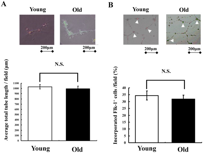
Figure 2. 3D culture of sorted Flk-1+ cells in vitro.
(A) Representative images of tube formation assay in vitro (upper). Sorted Flk-1+ cells derived from young and old iPS cells were cultured alone for 24 hours on Matrigel. Quantitative analysis of network projections formed on Matrigel for each experimental group (lower) (n = 3 in each group). (B) Representative images of HUVEC co-cultured with Flk-1+ cells (upper). Sorted Flk-1+ cells derived from young and old iPS cells were co-cultured with HUVEC for 24 hours on Matrigel. Flk-1+ cells derived from young and old iPS cells (white arrow head) were confirmed. The bar indicates 200 µm. Quantitative analysis of the number of Flk-1+ cells derived from young and old iPS cells into HUVEC on Matrigel (lower) (n = 3 in each group).

