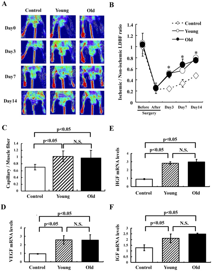
Figure 4. Effects of cell transplantation on blood flow recovery in the ischemic hindlimb.
(A) Representative LDBF images. A low perfusion signal (dark blue) was observed in the ischemic left hindlimb of control mice (PBS), whereas high perfusion signals (white to red) were detected in the ischemic left hindlimb of mice transplanted with Flk-1+ cells derived from young and old mice (2×10⁵ cells) on postoperative days 3, 7 and 14. (B) Quantitative analysis of the ischemic to non-ischemic limb LDBF ratio on pre- (Day-1) and postoperative days 0, 3, 7 and 14 (Control: n = 8, Young: n = 4, Old: n = 4). *p<0.05 for mice injected with Flk1+ cells (2×10⁵) vs. control mice. (C) Capillary density analysis. Capillary density was determined at day 21 after surgery. Collected ischemic hindlimb muscle was stained with VE-cadherin. Capillary density was calculated as below. The number of VE-cadherin positive cells per field was divided by the number of muscle fibers per field (n = 5 in each group). (D) VEGF, HGF and IGF synthesis in ischemic tissue determined by real-time PCR at day 7 after surgery following transplantation of Flk-1+ cells or PBS. VEGF, HGF or IGF mRNA levels were expressed relative to GAPDH mRNA levels (n = 5 in each group). N.S. = no significant difference between groups.

