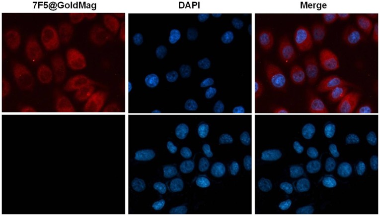
Figure 1. LSCM verifying specific targeting of 7F5@Au/Fe3O4 probe to PC-3 cells.
Red fluorescence (7F5@Au/Fe3O4rpar; observed in the membrane of the 7F5@Au/Fe3O4+ PC-3 cells (top row) while no red fluorescence was observed in the membrane of the 7F5@Au/Fe3O4+ SMMC-7721 cells (bottom row). Cell nuclei were stained blue in color via DAPI (middle column). 7F5@Au/Fe3O4 fluorescence images and DAPI images are merged in right-most column. Scale bar, 10 µm.

